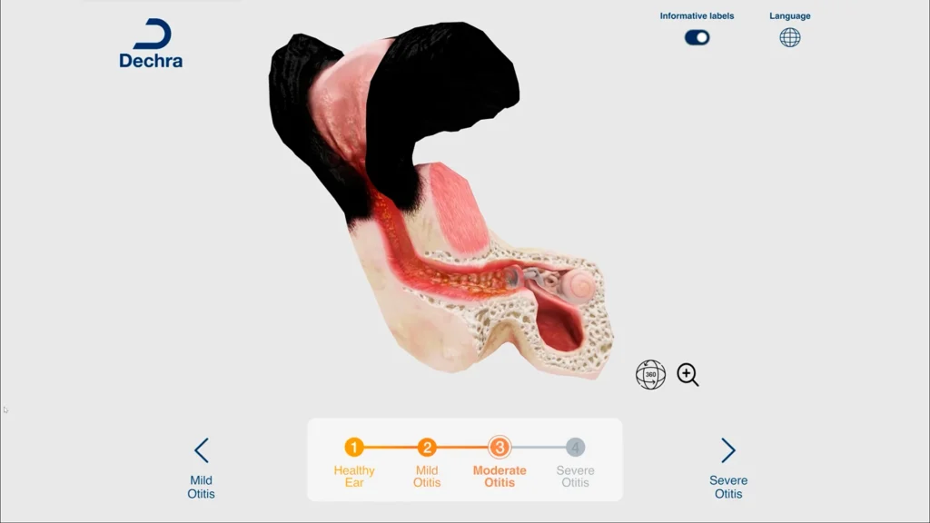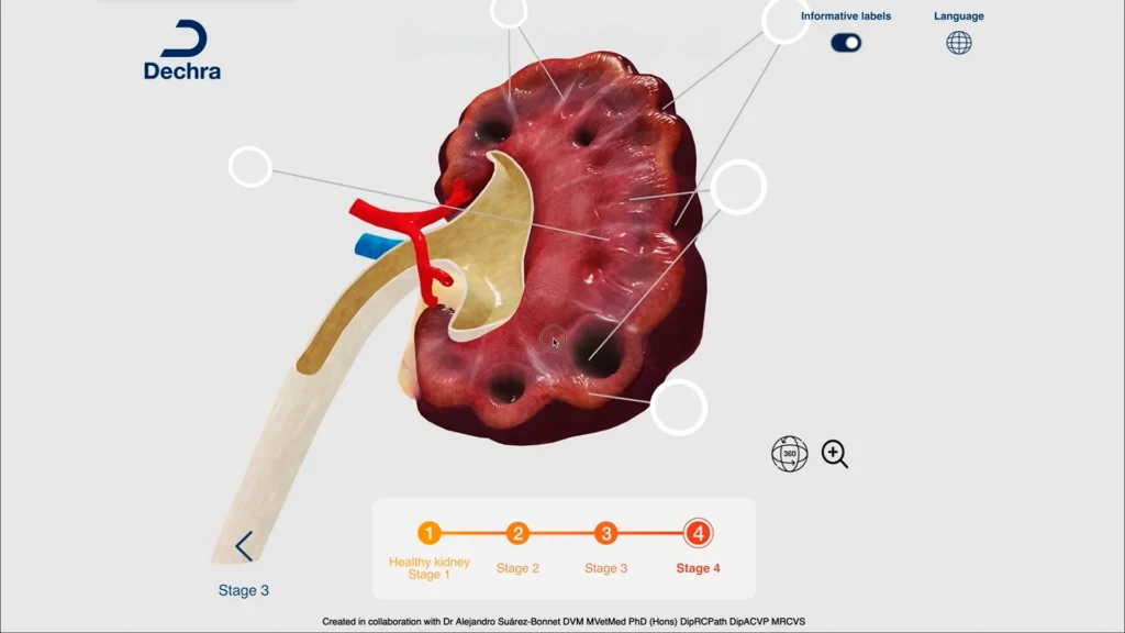
Dechra is a world-leading veterinary pharmaceutical company with commercial presence in 88 countries and more than 3,700 employees dedicated to developing medicines for animals. In a sector where ongoing training of healthcare professionals on animal pathologies is a recurring necessity, Dechra sought a tool that combined advanced scientific education and interactive visualization. The answer is Dechra 3D Anatomy, an interactive infographic featuring three-dimensional anatomical models of the kidney, ear and heart, which allows veterinary professionals to explore animal anatomy and observe the evolution of various pathologies across four stages.
01
The challenge
Teaching complex pathologies beyond the anatomical layers
Veterinary training in anatomy and animal pathologies has traditionally relied on static materials: illustrated slides, anatomical atlases, photographic sections, and masterly explanations. However, understanding a complex organ—its three-dimensional structure, its internal components, physiological variations, and the various pathological states it undergoes—is practically impossible with two-dimensional resources. Dechra needed a tool that would help healthcare professionals overcome this cognitive limitation: exploring the anatomy from within, understanding the mechanism of a pathology at each of its stages, and consolidating scientific knowledge through a visual experience that is natural and accessible.